Tendon Diagram - 2 814 Tendon Vector Images Free Royalty Free Tendon Vectors Depositphotos - This tendon connects the patella (kneecap) to the tibia.. Possibly the most important tendon in terms of mobility is the achilles tendon. Tendons are remarkably strong, having one of the highest tensile strengths found among soft tissues. The two peroneal tendons in the foot run side by side behind the outer ankle bone. Raises and rotates arm in all directions. There are over two dozen gorgeous and painstakingly detailed illustrations on this chart, from the extensor pollicis longus to the flexor digitorum.
Lower back muscle diagram anatomy does degenerative disc disease affect the lower back muscle? The knee joint is a complex structure that involves bones. The changes in ligaments and tendons generally occur more slowly than adaptation in bone, because ligaments and tendons have less vascular supply. This tendon connects the patella (kneecap) to the tibia. Also allows the action of raising up onto toes.
14 092 Tendon Stock Photos Pictures Royalty Free Images Istock from media.istockphoto.com Intermediate back muscles and c. Bones in shoulder, ligaments of the shoulder joint, parts of the shoulder joint, shoulder anatomy, shoulder joints and muscles, shoulder structure anatomy, shoulder tendon anatomy, shoulder tendons ligaments, human muscles, bones in shoulder, ligaments of the shoulder joint, parts of. The achilles tendon attaches the muscles of the calves to the bones of the ankle and foot. Raises heal when leg is straight. Tendons are the connection between bones and muscles. This tendon connects the patella (kneecap) to the tibia. It attaches to the wrist bone, the pisiform, and as well as the 5th hand bone. One peroneal tendon attaches to the outer part of the midfoot, while the other tendon runs under the foot and attaches near the inside of the arch.
The fleshy, thick part of the muscle is called its belly.
Ligaments join the knee bones and provide stability to the knee: Muscles and tendons of the human arm and hand, vintage engraved. Flexor tendon lacerations are classified into five zones 2, 15, 16. The two peroneal tendons in the foot run side by side behind the outer ankle bone. This diagram depicts muscle in the body 744×1054 with parts and labels. Extends spine and trunk back. Tendons are thick bands of tissue that connect muscles to bones. The fcu tendon is one of two tendons that bend the wrist. The achilles tendon enables us to walk, without it we would not be able to raise our heels of the ground. The achilles tendon is also called the calcaneal tendon. This sudden, tight, intense lower leg pain is sometimes called a charley horse. 9 photos of the foot tendons and ligaments diagram. The knee joint is a complex structure that involves bones.
This sudden, tight, intense lower leg pain is sometimes called a charley horse. The tendon travels along the inside of the forearm on the side of the small finger and crosses the wrist. Allows the action of raising the foot. On the other hand, the insertion is where a tendon attaches that muscle to the *more* movable bone. Intermediate back muscles and c.
Tendon Anatomy Anatomy Drawing Diagram from phase-iv.com On the other hand, the insertion is where a tendon attaches that muscle to the *more* movable bone. Muscles and tendons of the human arm and hand, vintage engraved. You can see a diagram of the achilles tendon below. The bones of the hip include the femur, the ilium, the ischium, and the pubis. Tendons are the connection between bones and muscles. The pubis, ischium, and ilium together constitute the pelvis while the thigh bone is the femur. This diagram depicts muscle in the body 744×1054 with parts and labels. The anterior cruciate ligament prevents the femur from sliding backward on the tibia (or the tibia sliding forward on the femur).
Tendon, tissue that attaches a muscle to other body parts, usually bones.tendons are the connective tissues that transmit the mechanical force of muscle contraction to the bones;
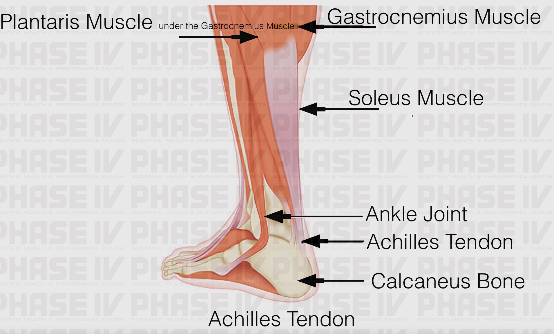
Attaches the calf muscles to the calcaneus, most important muscles for running, jumping, walking etc. Tendon, tissue that attaches a muscle to other body parts, usually bones.tendons are the connective tissues that transmit the mechanical force of muscle contraction to the bones; Bones in shoulder, ligaments of the shoulder joint, parts of the shoulder joint, shoulder anatomy, shoulder joints and muscles, shoulder structure anatomy, shoulder tendon anatomy, shoulder tendons ligaments, human muscles, bones in shoulder, ligaments of the shoulder joint, parts of. Bones, cartilage, ligaments, and tendons. The tendon travels along the inside of the forearm on the side of the small finger and crosses the wrist. This important tendon in the back of the calf and ankle connects the plantaris, gastrocnemius, and soleus muscles to. 2 ligaments (trapezoid& conoid ligaments) attach the clavicle coracoid process of scapula these tiny ligaments (w/ acominoclavicular joint) keep scapula attached to clavicle. Superficial posterior muscles of the forearm posterior compartment muscles of the forearm. The achilles tendon attaches the muscles of the calves to the bones of the ankle and foot. Diagram of tendons in forearm. Raises heal when leg is straight. Possibly the most important tendon in terms of mobility is the achilles tendon. Flexes elbow and moves forearm.
Lower back muscle diagram anatomy does degenerative disc disease affect the lower back muscle? Diagram of tendons in forearm. Fall on one point of shoulder and can rupture these ligaments with dislocation of ac joint. The fcu tendon is one of two tendons that bend the wrist. This tendon connects the patella (kneecap) to the tibia.
Tendon Structure And Function What Are The Tendons And What They Do Youtube from i.ytimg.com Flexes elbow and moves forearm. This diagram depicts muscle in the body 744×1054 with parts and labels. The achilles tendon is the largest. The two peroneal tendons in the foot run side by side behind the outer ankle bone. 17 photos of the diagram of shoulder muscles and tendons. The changes in ligaments and tendons generally occur more slowly than adaptation in bone, because ligaments and tendons have less vascular supply. Again, our knowledge of how mechanical stimulus mediates ligament and tendon structure is more empirical and less. There are over two dozen gorgeous and painstakingly detailed illustrations on this chart, from the extensor pollicis longus to the flexor digitorum.
Diagram of tendons in forearm.
Flexor tendon lacerations are classified into five zones 2, 15, 16. Foot anatomy diagram, foot joint diagram, foot sprain diagram, foot tendons and ligaments pain, leg tendon diagram. The changes in ligaments and tendons generally occur more slowly than adaptation in bone, because ligaments and tendons have less vascular supply. There are over two dozen gorgeous and painstakingly detailed illustrations on this chart, from the extensor pollicis longus to the flexor digitorum. The pubis, ischium, and ilium together constitute the pelvis while the thigh bone is the femur. 17 photos of the diagram of shoulder muscles and tendons. Allows the action of raising the foot. Tendons are thick bands of tissue that connect muscles to bones. In the back and elsewhere in the body, tendons attach muscles to bones. Ligaments and tendons are adapted in response to changes in mechanical stiffness. The bones together make up the hip. Brings leg back to and across body. This tendon connects the patella (kneecap) to the tibia.